 quoneon
Images and regions
Inspect CT and MRI, explore image histograms, masks, segmentation and spatial alignment.
Medical Image Processing
Move from medical images to quantitative research. Inspect volumes and regions, explore diffusion and perfusion, and examine spectroscopy with figures and numerical results that retain their scientific context.
117 apps in the catalog · Early access in preparation
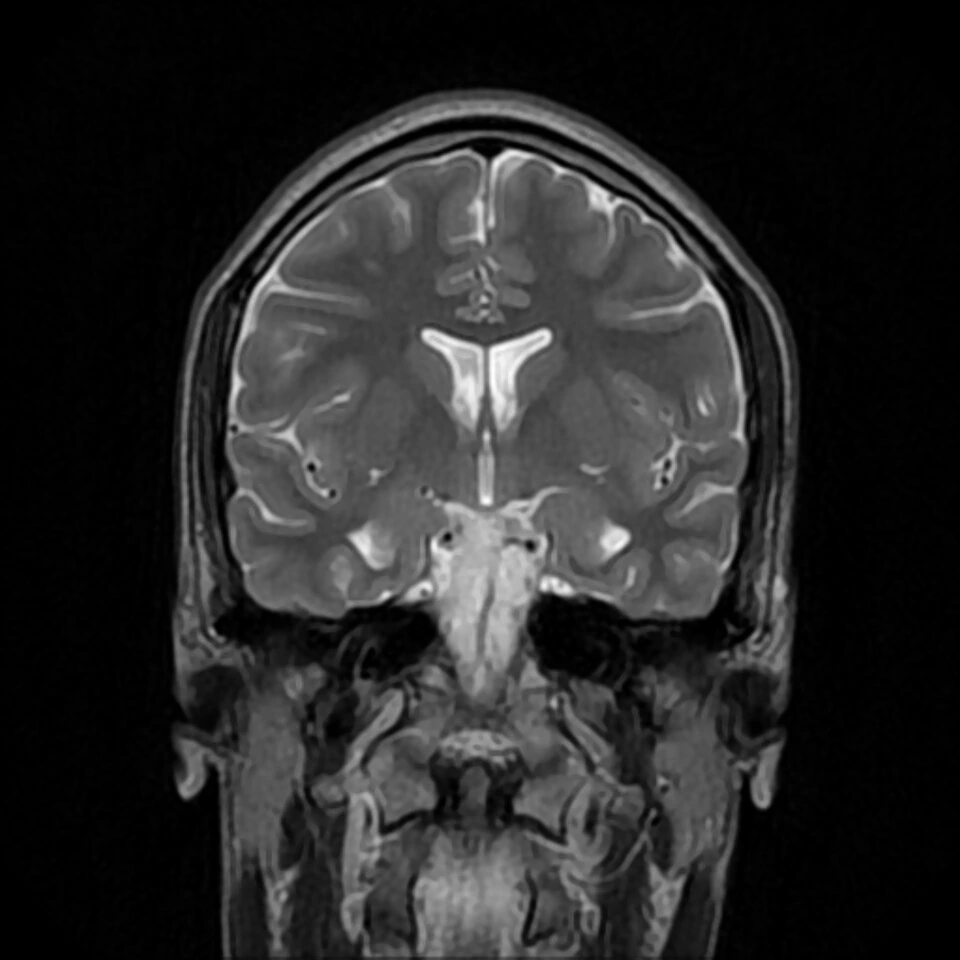
Inspect CT and MRI, explore image histograms, masks, segmentation and spatial alignment.
Work with diffusion measurements, tensor maps and supported tractography workflows.
Investigate time-dependent measurements and supported quantitative imaging models.
Explore spectral information and review complementary image sources.
Four apps from four areas, brought together in one narrated film.
Selected recorded app demonstrations and saved figures. Example data are identified on screen; these are research examples, not a live hosted session.
Supported image volumes, masks, acquisition metadata and numerical inputs.
Each workflow has its own required fields, units and supported formats. The guide helps you prepare the right input.
Image views, quantitative maps, regional measurements, spectra and supported result exports.
Read figures alongside the numerical values, settings and limitations of the selected analysis.
Follow a worked example, understand its inputs and inspect the results.
Your next research question
Tell us about your data and the workflow you want to use.